Ultrasound and Doppler Ultrasound and Doppler

Ultrasound and Doppler

Ultrasound scanning can help in almost any domain of disease identification.

About Ultrasound and Doppler

Ultrasound and Doppler

At Mahajan Imaging & Labs, we have highly trained ultrasound and doppler specialists working on the highest-end equipment available in the world today. We were the first to introduce 4D ultrasound scanning in India in 2004.

3D-4D Ultrasound

Mahajan Imaging & Labs is the leader in 3D-4D ultrasound

Mahajan Imaging & Labs has the unique capability to perform all routine and complex studies including FNAC’s, Ultrasound guided biopsies, Fibroscan with Shearwave Elastography, across our centres in an patient friendly and comfortable environment.

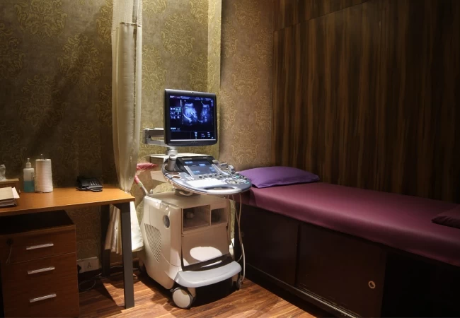
Ultrasound is the safest way to look inside the human body. It involves the use of high-frequency sound waves, which are harmless to the human body. Ultrasound scanning can help in almost any domain of disease identification. Ultrasound can be used to image many areas of the body including the pelvis and abdomen, the musculoskeletal system, the breast, the male reproductive system, the kidney, the thyroid, and salivary glands, the gall bladder, the pancreas and the developing foetus, liver, neck etc. Ultrasound is a valuable tool, and in the right hands, it can clear out most doubts related to the diagnostic queries that we are faced with.

Please note:

According to the PC-PNDT Act, performing a pregnancy ultrasound without a valid prescription and having the doctor’s DMC/HMC/MCI registration number, phone number and other details is forbidden.

General, Musculoskeletal and Vascular Ultrasound

Ultrasounds are simple, safe procedures that do not require the use of radiation to develop images of internal parts of the body. Ultrasounds can be used to examine many parts of the body, including the:

  • Blood vessels
  • Pelvis
  • Heart
  • Musculoskeletal system
  • Abdomen
  • Other soft tissue organs
Pregnancy Ultrasound

Ultrasound is a highly valuable diagnostic tool, and it is particularly useful during pregnancy because it is completely safe for you and your baby. Some of the useful indications for use during pregnancy include:

  • for dating purposes and to accurately determine your due date
  • to ascertain the number of babies present
  • to check for any bleeding early in the pregnancy
  • to check the position of the placenta
  • to assess the growth of the baby and its general well-being
  • to provide information about the anatomy of the baby and check for possible abnormalities*

*note all possible birth abnormalities are reliably diagnosed using ultrasound, and all scan results should be interpreted within the limitations of the test.

Shear Wave Fibrosis Scan

Shear Wave Elastography on Mahajan Imaging & Labs machines such as LOGIQ E9, Aplio i800, the most advanced machines globally, is the most advantageous technique for the non-invasive quantification of liver tissue stiffness.

Fibroscan (Liver Elastography)

Fibroscan is a non-invasive test that helps assess the health of your liver. Specifically, it uses ultrasound technology to determine the degree of fibrosis or scarring that may be present in your liver from various liver diseases or conditions. It is useful in patients with fatty liver in order to identify those with fibrosis who have progressive disease. It is also useful for the assessment of fibrosis and cirrhosis of the liver due to any cause.

Fibrosis Scan of the Liver by Shear Wave Elastography with Ultrasound
  • Quantitative measurement of tissue stiffness
    • Shear Wave speed (m/s)
    • Young’s Modulus (kPa)
  • 2D color-coded visualization of tissue stiffness
  • Image guidance with B-mode ultrasound images, which allows for:
    • Instant visual quality assessment of spatial stiffness, useful for detection and characterization of focal lesions
    • Better diagnostic interpretation with overlaying of shear wave stiffness images on the B-mode images
    • Optimal placement of Regions of Interest (ROI) for quantitative stiffness measures
    • Sampling within the ROI provides tissue stiffness measures at any location in the ROI, unlike conventional fibroscan which measures only the average stiffness within an ROI in a blind manner
  • Assessment and comparison of the degree of fibrosis in the same site of the liver on follow up imaging using this technique, which was not possible earlier.

Colour Doppler

Cutting Edge Technologies

Colour Doppler is a medical imaging technique utilized to offer visualization of the blood flow by using color processing to add color to the image so that a doctor can see what is happening inside the body.

A duplex ultrasound is a test to see how blood moves through your arteries and veins. The test combines traditional ultrasound with Doppler ultrasonography. Regular Ultrasound uses sound waves that bounce off of blood vessels to create pictures. Doppler looks at how sound waves are reflected off from moving objects, such as blood.

There are different types of duplex ultrasound exams. Some include:
  • Arterial and venous duplex ultrasound of the abdomen examines blood vessels and blood flow in the abdominal area.
  • Carotid duplex ultrasound looks at the carotid artery in the neck.
  • Duplex ultrasound of the extremities looks at the arms or legs.
  • Renal duplex ultrasound examines the kidneys.
Why It Is Done
  • Detect blood clots and blocked or narrowed blood vessels in almost any part of the body, especially in the neck, arms, and legs.
  • Evaluate abnormal veins causing varicose veins or other problems.
  • Find out the amount of blood flow to a transplanted kidney or liver.
  • Monitor the flow of blood following blood vessel surgery.

Available at:

Defence Colony

E -19, Ring Road, Defence Colony, New Delhi - 110024

Get Directions
Safdarjung Development Area (SDA)

C-6/8, Safdarjung Development Area, New Delhi – 110016

Get Directions
Pusa Road

7-B, Upper Ground Floor, Main Pusa Road, Rajinder Nagar, New Delhi - 110005

Get Directions
Gurugram

Ground Floor, Times Square Building, Sushant Lok Phase-I, Gurugram, Haryana - 122002

Get Directions
Fortis Hospital, Vasant Kunj

Fortis Hospital: Aruna Asaf Ali Marg, Pocket 1, Sector B, Vasant Kunj, New Delhi-110070

Get Directions
PSRI Hospital

Pushpawati Singhania Research Institute (PSRI): Pocket J, Sheikh Sarai Phase II, New Delhi-110017

Get Directions
Fortis Escorts Hospital, Jaipur

Fortis Escorts Hospital: Sector 5, Malviya Nagar, Jaipur, Rajasthan-302017

Get Directions
Text copied to clipboard