

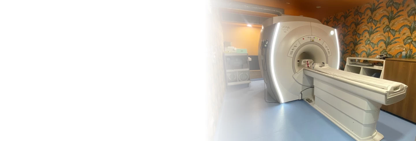
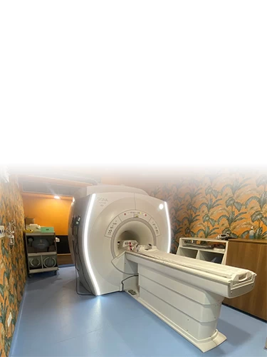
About 1.5T Volume MRI
In
1997, Mahajan Imaging & Labs introduced the first 1.5 Tesla MRI in a
stand-alone facility in India. Our diagnostic center is equipped with the
latest high-definition 1.5 Tesla MRI system from GE Healthcare, and all MR
examinations, including ultrafast sub-second scans, are routinely performed.
With the combination of up-to-date technology and a patient-centric environment, we make it easy for even anxious or pediatric patients to go through the MRI scan without any challenges.
Types of MRI
Functional MRI measures signal changes in the
brain that are due to changing neural activity and is also referred to as BOLD
imaging (blood oxygen level dependent).
A person’s brain is scanned while patient is
performing a certain physical task, such as squeezing a ball or looking at a particular
type of picture. When neural activity increases, oxygen demand increases too,
and the vascular system actually overcompensates for this, increasing the
amount of oxygenated haemoglobin relative to deoxygenated haemoglobin.
Such techniques are used in pre-surgical
planning and in basic neuroscience research in areas such as memory, expressive
and receptive speech, visual-spatial processing, and other cognitive processes.
Diffusion-weighted images are
very commonly used in the assessment of acute stroke and are also widely used
in oncology.
Diffusion Tensor Imaging, also known as MR Tractography non-invasively maps white matter tracts. Diffusion parallel to nerve fibers has been shown to be greater than diffusion in the perpendicular direction. This provides a powerful tool to study in vivo fiber connectivity in the brain in a noninvasive manner and preoperative planning of resectability of brain tumors. The application of this technique to stroke, Alzheimer's disease, and pediatric brain development is being investigated.
MR angiography is used to generate pictures of the arteries to detect any abnormal narrowing/dilatation or abnormal arterio-venous connections. MR angiography is often used to evaluate the arteries of the neck and brain, the thoracic and renal arteries, and the arteries of the legs. Magnetic Resonance Venography (MRV) is a similar procedure used to image veins.
MR spectroscopy is a noninvasive method for providing metabolic information about the brain. MR spectroscopy enables tissue characterization on a biochemical level that surpasses conventional MR imaging. MR spectroscopy also detects abnormalities that are invisible to conventional MRI because metabolic abnormalities often precede structural changes.
MR defecography is a technique for evaluating anorectal function. It is indicated in patients with constipation, rectal incontinence, painful defecation, and rectal prolapse. It permits analysis of the anorectal angle, the opening of the anal canal, the function of the puborectalis muscle, and the descent of the pelvic floor during defecation. It provides a clear visualization of the rectal wall, intussusception, enteroceles and rectoceles, along with an excellent demonstration of the perirectal soft tissue. It allows the assessment of spastic pelvic floor syndrome and descending perineum syndrome. No radiation hazard is associated with this procedure, unlike fluoroscopic X-ray defecography. Images are obtained with the patient at rest, at maximal sphincter contraction, during straining, and during defecation.
Although X-ray Mammography remains the primary imaging modality in the evaluation of breast disease, the mammogram maybe inconclusive for the presence or location of an abnormality. MR imaging has been used as an adjunct to mammography, particularly for patients with equivocal mammographic findings. The main advantage of breast MR imaging is its high sensitivity, with reported sensitivities for cancer ranging from 91% to 100% with varying specificity. Multiple investigators have shown that MR mammography may be useful to verify the multifocality and multicentricity of breast cancer, differentiating scars from recurrences after breast-conserving therapy, screening high-risk groups with a family history of breast cancer, investigating breast implants, examining breasts in cases of histologically proven breast cancer, and detecting metastases from an unknown primary.
Frequently Asked Questions (FAQ’S)
Certainly, a 1.5T MRI is generally safe for most people as it uses a magnetic field of 1.5 Tesla, which is considered to be of moderate strength. At Mahajan Imaging & Labs, we use advanced 1.5T MRI systems that harness a moderate 1.5 Tesla magnetic field, ensuring both high-quality imaging and strict adherence to safety protocols.
Typically, no special preparation is required for a 1.5T MRI. Although, it is essential to inform your healthcare professional about any allergies, health conditions or implants.
At Mahajan Imaging & Labs, duration may vary depending on the type of scan. However, a 1.5T MRI scan typically takes between 10 to 35 minutes.
Patients may experience a little discomfort due to lying still for a longer period. However, patients will generally not feel any pain.
In majority of cases, one can eat and drink before a 1.5T MRI scan without any specific restrictions. However, certain types of contrast enhanced scans or abdominal MRI may require fasting prior to the procedure. Consultation with a healthcare professional is advised.
Results for 1.5T MRI typically become available within 1 to 2 days after being diagnosed. However, the, duration may exceed depending on the urgency of the case and complexity of the images.
To assess scanning safely and conveniently, it is important to consult with your doctor beforehand as a pacemaker or metal implants may get affected by a magnetic field and malfunction during scanning. At Mahajan Imaging & Labs, our expert team carefully evaluates each case to ensure that the magnetic field will not interfere with your implants, ensuring a safe and effective scanning experience.