

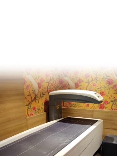
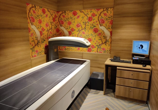
About Dexa Bone Densitometry
Bone densitometry is a type of X-ray used to measure the
bone mineral
density. It is a simple, quick, and
non-invasive medical test that involves exposing targeted
areas like the spine or hip to a very small amount of
ionization radiation.
DEXA bone densitometry is used
to assess bone mineral density in specific target areas or the entire body, and
it is especially beneficial in postmenopausal women and the elderly.
The test helps healthcare experts know if the bone mass has decreased, as it makes the bones more brittle and increases the chances of a fracture.
Osteoporosis is a metabolic disease affecting the skeleton which causes a reduction in the amount of bony tissue. Bones are weakened as these tissues are reabsorbed or taken up by local cells. At the core, bones become less dense, on the perimeter, cortical bones lose thickness. Complications from osteoporosis arise as bones become thinner, more porous, and more susceptible to fractures.
Usually occurs in women during menopause. At this time, the ovaries produce less estrogen, a female sex hormone. In the absence of estrogen, bone reabsorption decreases, dropping overall bone mass below the maintenance density level, leading to a high risk of fractures.
Age-Related Osteoporosis – Inflicts both women and men older than 70 years. Older people have an added risk of low bone mass because bone density peaks at the age of 35 and decreases gradually. The ability to absorb calcium from the intestine decreases, thus reducing the calcium inside the body. Also, older people are slightly vitamin D deficient, leading to decreased calcium absorption from the intestine. Bone formation responds to physical stress, and thus, less activity also decreases bone strength.
Osteoporosis may
go unnoticed if it is asymptomatic. Signs that there has been a reduction in bone
mass include:
Loss of teeth and
height over time is often accompanied by a stooped posture. Minimal trauma
fractures, i.e., fractures occurring without the application of significant
force, as bone density decreases, the risk of fracture increases.
Frequently Asked Questions (FAQ’S)
A DEXA scan is performed to assess the risk of fractures and measure bone density. A DEXA scan can effectively diagnose conditions like osteoporosis, monitor bone health over time, and helps guide treatment decisions. The scan is fairly quick, non-invasive, and provides accurate measurements of bone strength and density using low radiation.
DEXA scan provides more detailed, accurate measurements of bone density, distinguishing it from other bone density tests. Unlike X-ray tests or ultrasound, DEXA uses low-dose X-rays to assess bone strength at specific sites like the spine and hip, offering a more precise evaluation of bone health.
Duration for a DEXA scan typically takes approx. 10-15 minutes on table.
A DEXA scan is highly recommended in postmenopausal women, older adults, those at the risk of osteoporosis, or those on medications that may affect bone health. A DEXA scan also assesses body composition in athletes and monitor bone health during treatment. At Mahajan Imaging & Labs, our experienced team helps the patient determine if a DEXA scan is appropriate for their specific needs.
A DEXA scan does not come under annual health check-up packages. Generally, it’s recommended every 1-2 years for those already diagnosed with osteoporosis or at a high risk for the same. In other cases, an individual's healthcare provider may suggest scanning every 2 to 5 years, depending on the risk factors and the individual's clinical health.
A DEXA scan is considered very safe, with only a small amount of radiation involved, much less than a standard X-ray, classifying it as a low-risk procedure. Its radiation exposure is quite minimal and is not considered harmful to most people.
Yes, one can eat and drink prior to a DEXA scan. However, it is recommended to avoid calcium supplements before scanning as they can affect the results.
Certainly, there are alternatives to a DEXA scan for measuring bone density:
- Ultrasound: Bone density is measured using sound waves, primarily for screening purposes.
- CT scan (Quantitative Computed Tomography): A 3D image is used to assess bone density, however, it exposes patients to more radiation compared to a DEXA scan.
- X-ray: While standard X-rays can detect fractures or bone loss, they lack the precision offered by DEXA scan in measuring bone density.