
About CBCT/OPG
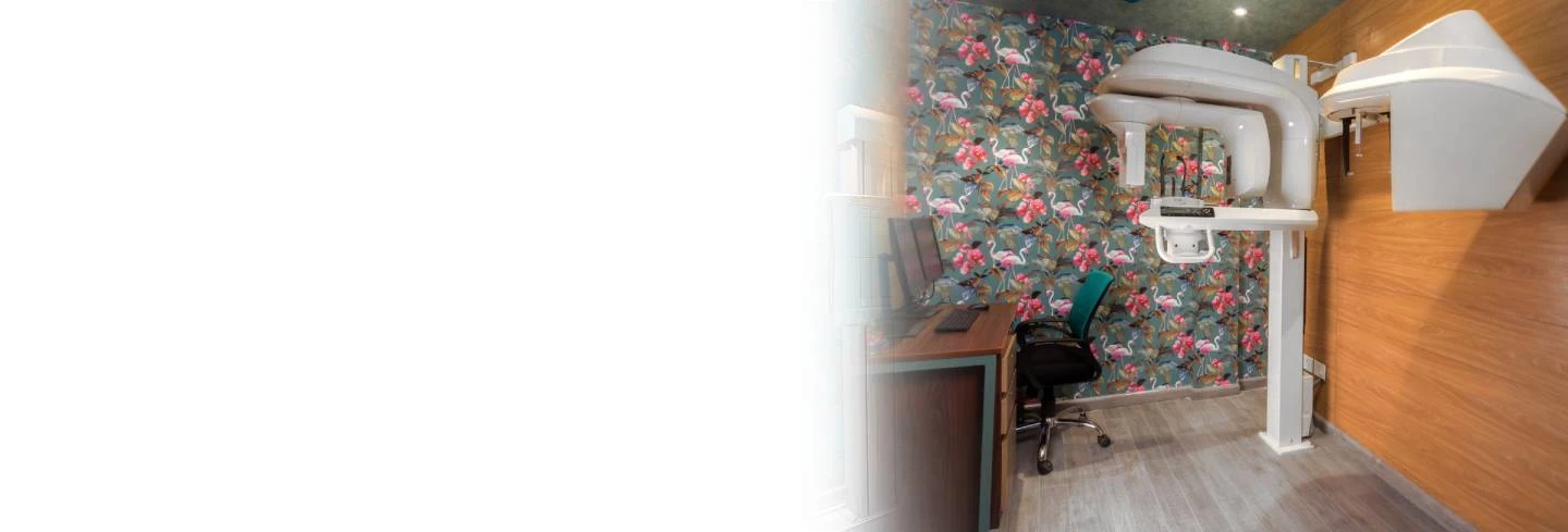
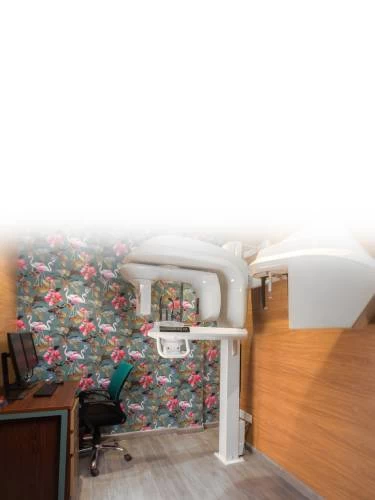
Cone Beam Dental CT (CBCT): A dedicated CT scanner for maxillofacial and dental applications region.
The Cone Beam CT uses an amorphous silicon flat panel detector and cone beam technology to get volumetric images with vastly lower radiation dose and higher precision when compared to traditional 16 or 64 slice Ct Scanner. This new age device provides a full range of diagnostic views including complete scans of all oral and maxillofacial structures, paranasal sinuses and airways. Acquired information gives the treating doctor more thorough structural knowledge which enables highly accurate diagnosis and treatment planning and can help create more predictable outcomes. Vastly lower radiation dose (20-100 times) means that the CBCT scan can be done safely without any undue concern for radiation side-effects.
What is Cone Beam Computed Tomography (CBCT)?
Computed tomography has been expanding rapidly and CBCT is the latest, most advanced generation of CTs. Cone Beam technology uses a cone shaped X-Ray beam that projects onto an amorphous silicon flat panel detector. The scanner rotates 360 degrees around a patient's head in a matter of seconds with the patient in a comfortable sitting position. The single turn motion image capture used in Cone Beam CT is quicker than conventional spiral motion, and can be accomplished at a lower radiation dose as a result of no overlap of slices. Cone beam CT provides views that can be presented as 3D volumes or 2D images for diagnosis and advanced planning.
Why the need for CBCT?
Traditional multi slice (6, 8, 16,32,40,64,128,256 and 320) CT scanners have been used in the past for dental and maxillofacial imaging. At Mahajan Imaging & Labs, we introduced DENTASCAN in Feb 2004 which provides similar 3D volume and 2D images.
Very Low Radiation Dose: The CBCT gives a radiation dose of about 36~Sv as compared to 2000~Sv to 4000~Sv of multi-slice CT.
Higher Precision and Accuracy: The CBCT provides images of superb clarity and great precision. What you see is real and the measurements are very accurate.
Dedicated Scanner: The CBCT is a dedicated scanner designed specifically for dental and maxillofacial imaging.
Dedicated Software: The dedicated user-friendly software simplifies treatment planning and provides necessary perspective in many cases. True-size, distortion free, high-resolution images are reconstructed rapidly, tailor-made for the needs of dental and maxillofacial professionals.
Eliminates Need for Multiple Exposures: The CBCT provides all information in one scan including panoramic and cephalometric images and 3D volumes, virtually eliminating the need for conventional orthodontic X-Rays. Patients can be comforted by the knowledge that the doctor will have all the information needed to evaluate their problem and plan treatment.
The Uses of CBCT
- Impaction/ Localization of teeth or Foreign body
- Oral surgery/Third Molar inerve relationship
- Orthodontics
- Pre-Implant imaging/Placement/Planning
- Intra-bony pathology
- TMJ Studies
- Asymmetry studies
- Orthognathic surgery
- Cosmetic surgical planning
- Endodontic canal assessment
- Airway evaluation
- Para nasal sinus investigation
Who will provide the report?
At Mahajan Imaging & Labs, a trained and qualified super specialist dental surgeon would read the scans and provide the report along with the films. Images and data would also be provided on a CD so that the referring doctor can also view the same on his own computer.
With volume rendering, clinicians can easily manipulate, enhance, and slice the volume in any orientation or shape for quick and effective diagnosis. Volume rendering is an effective tool for surveying skeletal morphology, dentition, and is a powerful patient demonstration / communication method.
Clinicians who desire a more in-depth investigation of internal structures can examine slice by slice and sweep through the entire volume using section views. This enables more precise evaluation/measurement of anatomical structures such as assessing the number/shape of root canals, TMJ characteristics, alveolar ridge thickness &: anatomy, airway/sinus, bone density/ defects, Hounsfield unit calibration, nerve pathway ID &: marking etc.
Tru-Pan delivers anatomically accurate and precise panoramic images with optimal clarity and detail. Tru- Pan's advanced reconstruction of unique anatomic landmarks automatically creates an individualized focal trough specific to each patient. The automatic custom focal arch detection works in conjunction with the patient's 3-D data to quickly and efficiently extrapolate "true" and precise panoramic views. There is no image distortion which allows for true measurements.
This feature gives clinicians the ability to easily and quickly construct and view traditional lateral and AP cephalometric radiographs, take measurements, and utilize several image enhancement features which dramatically increase the viewing options allow easier viewing of landmarks.
Clinicians can perform image based planning for restorative implants or orthodontic mini screws using the implant planning feature. This feature enables precise implant planning through simultaneous buccal, lingual, vertical, and density visualization. The tool offers straightforward analysis within brand of implant.
The CBCT Scan generates a limitless number of x-rays India from one scan. Linear and angular measurements can be made on any of the acquired images. Standard examples are coronal and sagittal TMJ slices, bitewings, PA's, maxillary occlusal views, mandibular occlusal views, etc.
Assessing treatment outcomes with the 3D superimposition of CBCT volume data is ground breaking. Our facility allows two CBCT scans to be opened at the same time in 3D and to be superimposed based on registering common stable landmarks on each scan.
The result is a 3D volume rendering of both scans with different colouring to highlight the difference between the scans.
- Orthodontic Diagnostic Assessment
- Implant Planning
- Tru-PAN OPG
- Orthognathic Surgical Evaluation
- Tooth & Alveolar Bone Evaluation
- CBCTscan Maxilla & Mand
- CBCTscan Face
- CBCTscan TM Joints
- Dual CBCTScan for guided implant surgery
- Implant Planning and Simulation
- Facilitation of Surgical Guide Fabrication
- Digital Orthopantomogram (OPG)
- Digital Cephalogram
- MRI TM Joints
- Contrast Enhanced MRI Face and Neck
- Non Contrast Parotid and Submandlbular MR Sialography
- Contrast Enhanced CT Face and Neck Parotid and Submandibular Sialography