

About 3T Digital MRI
This latest generation 3.0T Digital MRI machine ensures fast and accurate MRI imaging. The specially designed ambient MRI room along with the latest silent MRI technology, which reduces sound levels, truly maximizes patient comfort. The standard applications portfolio contains imaging solutions that cover a wide variety of contrasts, 2D and 3D volumetric data, including motion correction capabilities.
Patient-centric MR Experience:
We have taken utmost care in all aspects to ensure a positive diagnostic imaging experience for every patient, be it adult or child.
One of the most common reasons why patients become uncomfortable and sometimes frightened during an MRI scan is due to the noise produced by the scanner. Conventional 3.0T MRI scanners generate noise up to 120dBA (similar to thunder / race car/ drilling machine). At Mahajan Imaging & Labs, the 3.0T MRI system features ‘Silent MRI’, resulting in only ambient room noise. This means, absolutely no noise during scanning of certain anomalies and maximum patient comfort and a relaxed experience.One of the most common reasons why patients become uncomfortable and sometimes frightened during an MRI scan is due to the noise produced by the scanner. Conventional 3.0T MRI scanners generate noise up to 120dBA (similar to thunder / race car/ drilling machine). At Mahajan Imaging & Labs, the 3.0T MRI system features ‘Silent MRI’, resulting in only ambient room noise. This means, absolutely no noise during scanning of certain anomalies and maximum patient comfort and a relaxed experience.
Patients with claustrophobia are very uncomfortable and anxious while entering the MRI tunnel in a routine MRI machine. However, the 70 cm wide-bore tunnel, with bright inner tunnel light and ventilation system in our 3.0T MRI ensures patient comfort and helps them overcome anxiety.
Our machines are equipped with unique in-bore audio-visual features where claustrophobic patients can watch TV while their scan is going on.
In conventional MRI machines, patients are required to enter the MRI tunnel with their head first, causing discomfort to some patients. In our MRI systems, the feet-first imaging for all body parts (including brain) can be performed i.e., the patient’s feet will enter the MR tunnel first, thus allowing an unrestricted view of the ambient surroundings and making patients feel more comfortable.
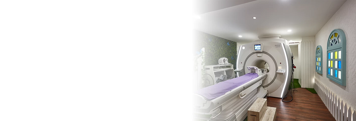
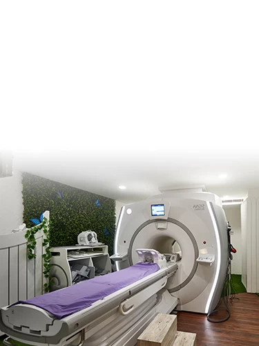
The sight of an MRI system can be overpowering for patients when they walk into a sterile MRI room for the first time and see a large MRI system sitting inside. Keeping this in mind, we have designed the scan room environment to mimic the calm serenity of a natural landscape, ensuring patients are comfortable and relaxed while being scanned, a first of its kind in the world!
The True Power of 3.0T
The biggest challenge with
conventional 3.0T MRI systems is dielectric shadowing artifacts (image
distortion), distorting images, and thus interfering in the accuracy of
diagnosis, thereby defeating the purpose of having MRI systems with higher
magnet strength. With the latest Multidrive RF Transmit technology, our DIGITAL
3.0T system automatically delivers uniform signal quality over a broad range of
patient sizes, eliminating the previous challenges of conventional 3.0T MRI,
ensuring consistently clear and distortion-free images.
The conventional 3.0T MRI systems
acquire the patient data with coils and transmit them to an electronics room
through copper cables. In our DIGITAL 3.0T MRI system, the signals are
digitized in the machine room itself and then transmitted to the electronics
room via Broadband Optical Fiber Cable technology, thus minimizing signal loss
and improving the image quality for better diagnostic confidence.
Our technologically superior 3.0T
machine overcomes tissue heating by automatically customizing and delivering
the optimal radiofrequency pulses as per the size and shape of the patient,
through its personalized patient management system.
Our latest generation 3.0T offers clinical benefits across spectrum of medical specialties by using advanced applications in Neuro, Spine, Vascular, Abdomen, Whole-body, Breast, Cardiac, Musculoskeletal, Orthopedic, and have espoused the new horizons in diagnostic capabilities and have the potential of change in course of treatment.
48 Channel Head Coil – Neuro Imaging made simple and perfect
In order to provide most accurate
and high-quality diagnosis related to neuroradiological imaging (i.e., for
Tumors, Stroke, Dementia, Infection, Aneurysms, Seizures, Cancer detection
etc.), a high-end sophisticated machine with higher number of channels is
needed. Our 3.0T MRI system comes with a 48-channel head coil, one of the best
coils available today globally. With this, neuro-radiological imaging has
become simpler and shorter than never before, yet offering highest resolution
brain imaging.
Now even the most advanced clinical exams like functional Imaging of Brain (fMRI – including the stimulus and response setup - visual, auditory, olfactory, gustatory, tactile, motor, cognitive) can be performed at ease and with utmost accuracy to understand the brain function and differences in various disease states.
Other Advanced Clinical Applications
Functional Nerve Imaging is direct imaging of the nerves in the body to look for pain points / compression. This technique yields a detailed image of a nerve from the diffusion signal that arises from the nerve itself rather than from surrounding tissues or from fat in the nerve lining. This is especially useful for the evaluation of the brachial plexus, lumbosacral plexus, sciatic nerve and pudendal nerve amongst others.
A boon for patients with renal disease who cannot be given contrast agents, we have the capability to image all the major arteries and veins of the body without any injection. This is particularly useful in patients with peripheral artery disease, varicose veins etc.
Mavrik enables us to scan patients with metallic implants. Previously, MRI machines were not able to clearly view areas of the body (knees, hips etc.) with metal implants, but with MAVRIK sequences that is now possible!
Advanced Neuro Applications suite includes SWAN, 3D COSMIC, Diffusion Tensor Imaging, Fibertrak, Brain Stat (AIF parametric maps), BrainWave Real Time, BrainWave Fusion etc., for clinically advanced neuro imaging and accurate assessments which were previously a huge challenge in MRI.
With this technique, both qualitative and quantitative information of the fat content in Liver can be obtained in a single breath hold. Liver fat content assessment is important for: Non-Alcoholic Fatty Liver Disease (NAFLD), Alcoholic Liver Disease (ALD), surgical planning, donor evaluation, and nodule assessment.
Brain perfusion assessment is important for Neuro Developmental Delay (NDD), tumors, stroke/TIA, AVM etc. The DSC Perfusion is not always reproducible & provides relative perfusion only. Whole-brain, quantitative tissue perfusion, without contrast, is great for the patient and is much simpler than injecting contrast.
Frequently Asked Questions (FAQ’S)
Certainly, a 3T digital MRI is considered safe. However, it is usually not recommended for individuals with certain medical conditions or implants as it creates detailed images of the body using a strong magnetic field and radio waves.
A 3T digital MRI's high resolution allows detailed imaging of the tissues and organs that helps diagnose multiple conditions, such as brain disorders (strokes and tumours), joint injuries, spinal issues, soft tissue damage, cancer, and multiple sclerosis.
At Mahajan Imaging & Labs, the scan duration may vary depending on its type. However, a 3T digital MRI scan typically takes between 10 to 35 minutes. This depends on the complexity of the procedure and the area being scanned.
Typically, no special preparation is required for a 3T digital MRI. However, some scans may need fasting for a contrast dye injection. It is required to consult a healthcare provider or the center before appearing for the examination.
A referral from the patient’s health care provider is required. An appointment for 3T digital MRI can be conveniently booked at Mahajan Imaging & Labs through our official website www.mahajanimaging.com, by calling our customer care number at +91 11 4118 3838 (from 7:00 A.M. to 10:30 P.M.), or chat with our WhatsApp bot at +91 88828 97661.
At Mahajan Imaging & Labs, results are typically available within 24 to 30 working hours after the patient’s scan, ensuring a prompt review.
Patients may experience little discomfort due to lying still for a longer period of time. However, patients will generally not feel any pain.
The key difference lies in the strength of the magnetic field. A 3T digital MRI uses a 3-tesla magnet, which produces images with higher resolution and greater clarity than standard 1.5T MRI machines. This enhanced imaging capability can detect smaller abnormalities, potentially leading to earlier diagnosis and improved treatment planning. At Mahajan Imaging & Labs, we leverage this advanced technology to provide precise and reliable diagnostic imaging.