


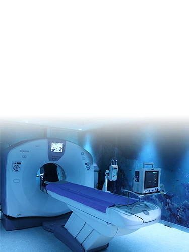
About 128/64/16 Slice CT
(Non-invasive CT Angiography)
Mahajan Imaging
& Labs is one of the first to introduce 16, 32, 64, and 128 Slice CT
scanners. The advanced scanning technologies, combined with our CT specialists,
are able to provide the state-of-the-art for diagnosing all types of illnesses
in the body. These scanners are capable of performing cutting-edge tests like
Coronary Angiograms (Heart Scans), Pulmonary Angiograms, Brain Angiograms,
True-match imaging, etc.
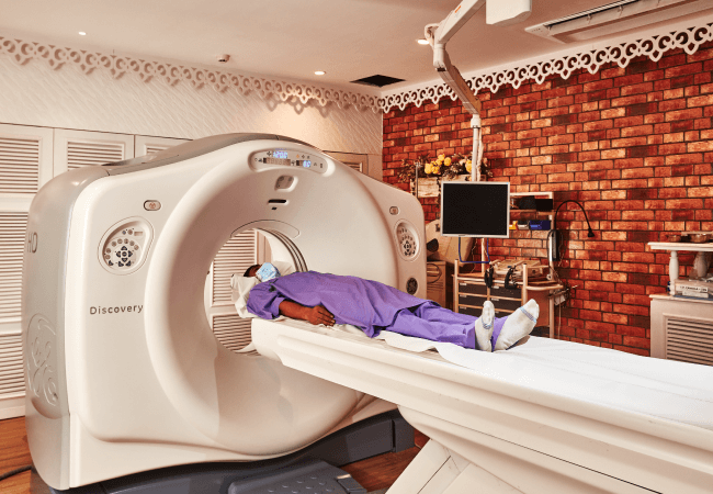
We were amongst the first to introduce multi-detector CT into the country and to perform non-invasive CT coronary angiography. Our group has multiple CT scanners, ranging from spiral to the highest end multi-detector CTs. The state-of-the-art 64 detector row (500) CTs provide ultra-fast volume imaging with the capability of a five-beat heart scan. Further, all routine CT scans are done using ultra-thin 0.625mm slices with volume acquisition and isometric reconstruction that can be obtained in any plane. 3D-CT examinations are routinely performed along with a virtual colonoscopy, virtual bronchoscopy, and whole-body scanning. Patients pay the same charges for routine CT scans as they would at any other facility that does not have these high-quality machines.
Non-invasive
imaging technologies continue to revolutionize every subspecialty of medicine.
This scanner has a special X-ray tube and rotation speed, making it capable of performing very rapid scanning with 64 detector rows. As per the recent US-FDA documentation based on the image acquisition criteria, this can be called a 440 slice/second CT. It is used for performing non-invasive CT angiograms of the heart, brain, and other blood vessels of the body. The 64-channel configuration also provides breakthrough performance in advanced pulmonary imaging, multi-organ trauma evaluation, and low-dose paediatric applications to boost clinical capabilities to the highest level attainable.
16 Slice CT Scanner
Mahajan Imaging & Labs latest 16 Slice CT
provides the benefits of high-resolution, low dose scanning with increased
integration and collaboration. It provides consistent image quality across a
diverse patient population and a wide range of exam types, supporting diverse diagnostic needs.
This high-end CT enables us to
do scans in one go of the neck, chest and abdomen. Also, we can do triple-phase
studies of the abdomen with submillimetre scan thickness. This state-of-the-art
CT machine is capable of doing high-resolution CT Angio of the abdomen down to
the feet in one acquisition and at a lower contrast dose. Other applications
include brain perfusion, lung nodule analysis, and vessel analysis, allowing
you to care for a wide range of patients with ease and efficiency.
The contrast in an image is crucial in differentiating between a lesion and a normal parenchyma or a normal structure. The latest technology embedded enhances the differentiation between a lesion and normal parenchyma, it helps identify lesions that are very iso-dense or iso-attenuating which would otherwise be difficult to differentiate from normal parenchyma. Multiple advanced applications on the console allow for post-processing without a stand-alone workstation.
- Enhance diagnostic confidence with superb image quality
- See more detail across a range of patient types
- Provide exceptional image quality for even small patients
- Streamline the workflow and reduce variability
The 70 kV scan mode – the first in a system of its kind – helps take patient care to a new level by offering low-dose scanning of smaller patients and allowing for protection of radiation-sensitive organs. This scan mode offers up to 20% lower dose scanning than 80 kV.
The award-winning technology found in our CT systems is one of the ways that CT is able to offer such excellent and exceptional image quality at low doses. It helps personalize image quality based on patient needs at a low dose and improves image quality through artefact prevention and increased spatial resolution at a low dose. It also allows for improved tube efficiency.
Frequently Asked Questions (FAQ’S)
The difference between a 128-slice, 64-slice, and 16-slice CT scan lies in the number of X-ray slices captured during each rotation. Higher slices ensure quicker scans and better image resolution. A 128-slice scan offers highest detail and speed compared to a 16-slice scan that offers lower resolution and slower imaging.
Typically, no special preparation is required for a 128/64/16 slice CT scan. However, some scans may need fasting for contrast dye. At Mahajan Imaging & Labs, we recommend consulting with your healthcare provider, or the ones at our our center, beforehand to ensure you follow only specific instructions.
Generally, 128/64/16-slice CT scans are safe, with minimal exposure towards radiation. Although some patients may get affected in kidney functioning, especially those with pre-existing kidney conditions, and experience allergic reactions from contrast dyes. Under certain conditions, consultation with patient's healthcare provider is highly recommended, for a convenient examination.
At Mahajan Imaging & Labs, results for a 128/64/16 Slice CT Scan typically become available within 24 to 30 working hours of the scan being done.
Certainly, children can undergo a 128/64/16-slice CT scan as we ensure special precautions to minimize radiation exposure. Although, the scan is only recommended when it is absolutely needed for the diagnosis.
A higher slice count enhances image quality by delivering finer and more detailed cross-sectional images of the body. This allows in reducing blurring, and for clearer visualization of structures enhances the ability to detect small abnormalities.
A 128/64/16-slice CT scan can diagnose multiple conditions and diseases including conditions such as internal bleeding and fractures, vascular conditions, infections, cancer, and certain lung diseases (emphysema, pulmonary embolism).
Generally, a CT scan is avoided during pregnancy unless it is absolutely necessary as high risks are associated with the radiation on the developing foetus. If medical implants are present, it’s important that your doctor is informed, as some implants may be affected by the procedure, though most are considered safe during a CT scan. At Mahajan Imaging & Labs, we adhere to strict safety protocols and take extra precautions to ensure the well-being of all our patients.